Overview of Virus

What is virus?

A virus (from the Latin virus meaning toxin or poison) is a small infectious agent that can replicate only inside the cells of other organisms. All virus contain a set of genes which is either RNA(Ribonucleic acid) or DNA (Deoxyribonucleic acid), a protein coat called capsid protein that protects these genes. Some virus also contains a viral envelope around the whole capsid protein. Viruses vary from simple helical and icosahedral shapes, to more complex structures. Most viruses are about one hundred times smaller than an average bacterium.


http://en.wikivisual.com/images/e/eb/Icosahedron.jpg
http://www.mcb.uct.ac.za/tutorial/virovirion.jpg


The picture below will explain how the virus works.

Example shown below will be done on Influenza virus.

Taken from Times magazine August 24, 2009.

Relationship between microbes and host.

There are three types of relationship between microbes and host.

  • 'Normal' microflora
  • Virus Host Relationship

'Normal Microflora'

Human babies are free from microbes in utero while lactobacilli in the mother’s vagina. There are many sources of flora in newborns babies and some of the examples are their families, personal or family doctors and those people who had physical contact with the babies. In a healthy individual, the internal tissues are free from microbes but the surface tissues are usually colonized by microbial species and the most common is bacteria, or sometimes fungi.


However, there are 2 views of viral flora in humans.
  • Yes, able to isolate viruses from seemingly 'healthy' people
  • No, these 'healthy' people are already immuno-compromised

Virus-Human Relationship

There are three types of virus-human relationship.

  • Commensalism – a class of relationship between two organisms where one organism benefits but the other is unaffected.


  • Mutualism - is a biological interaction between two organisms, where each individual derives a fitness benefit (i.e. increased survivorship). Similar interactions within a species are known as cooperation.


  • Parasitism – Parasitism is a type of symbiotic relationship between organisms of different species where one organism, the parasite, benefits at the expense of the host.

Portals of Entry

Portals of Entry

Virus enters host into 4 different sites:

  • Skin
  • Mucous membrane
  • Placenta
  • Parental route

Pathogens can cross placenta and infect foetus which leads to birth defect, premature birth and spontaneous abortion. In the skin, it is barrier to most pathogen. Skin infections are more frequent in immunodeficient patients, seniors and infants than in healthy individuals with an intact immune system. The severity of infections ranges from localized, self-limiting cutaneous infections to widespread necrosis of the skin, muscle, and fascia. The most frequent infections of the skin and mucous membranes are caused by bacterial and viral infectious agents. For parental route, pathogens are deposited via punctures like nail and needles.

Types of General Virus Replication Cycle

There are 2 types of General Virus Replication Cycle.
  • Lytic cycle
  • Lysogenic cycle
The picture below will show a more indepth phases of these 2 cycles.


http://textbookofbacteriology.net/themicrobialworld/lysogeny.jpg

Phases of General Virus Replication Cycle

There are 6 phases in a General Virus Replication Cycle, they are

  1. Attachment
  2. Penetration
  3. Uncoating
  4. Replication and Expression
  5. Maturation
  6. Release

http://www.influenzareport.com/ir/images/image26.jpg

1. Attachment

All viruses contain one more important proteins known as the attachment proteins or docking proteins. The attachment protein is needed by the virus to attach to its target cell before it can enter that cell. Attachment proteins lie on the outer surface of the virus and contact the appropriate receptor sites on the target host cells, in other words, receptor sites are specific. Receptors on host cells may be protein, glyroprotein or glycolipid. These attachment proteins are often called spikes because they can extend away from the cell so as to better be able to contact the host receptor (think of the viral AP & the host receptor site as being the pairs of a velcro systemVELCRO SYSTEM). In addition, virus may contain small quantities of carbohydrate (glycoprotein). Cells lacking in specifics proteins are resistant to virus infection.


2. Penetration

Penetration follows right after attachment process. There are 3 ways of penetration, namely, receptor-mediated endocytosis (enveloped virus), clathrin-mediated endocytosis (naked virus) and fusion for eukaryotic cells. Endocytosis is the process whereby cell absorbs molecules from outside the cell by engulfing them with their cell membrane. In the case of receptor-mediated endocytosis, enveloped virus contains attachment proteins which bind to the host’s receptor site, after binding, the cell membrane will engulf the virus into cell, thus successfully penetrating the cell. As for clathrin-mediated endocytosis, it can be shown from the animation below.

http://stke.sciencemag.org/content/vol2004/issue264/images/data/re19/DC2/slowtrack2.swf

As for fusion, the envelope of virus fuses with the cell membrane as the virus approaches the cell membrane, thus releasing the virus nucleic acid into the cell.

For prokaryotic cells, the phage binding to the host cell receptor sites through its docking proteins at the end of the tail fibres. Once firmly attached to the cell, a viral enzyme in the tail punches a hole in the host's cell wall and the core region of the tail is thrust through and the DNA ejected into the host's cytoplasm. In the case of phage the protein coat remains on the outside, but with eukaryotic cells the entire virus may be taken into the host's cytoplasm.


3. Uncoating ( only in animal viruses)

Uncoating is the process whereby the nucleic acid is separated from its protein coat. This is done so by the digestive enzymes, lysozymes, produced in the host lysosomes. The viral enzymes produced by the virus itself, also separates the nucleic acid from the protein coat.
They are many types of uncoating,

  • Pore formation: picornavirus
  • Direct plasma membrane fusion: Paramyxoviridae
  • Partial plasma membrane fusion: Human Immunodeficiency Virus (HIV)
  • Endosomal plasma membrane fusion: influenza virus
  • Endosomal plasma membrane lysis: adenovirus


4. Replication & Expression

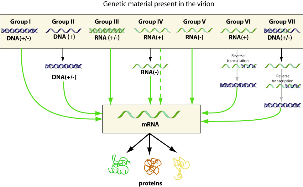
Once inside the host cell, the viral genome takes over the metabolism of the host, converting the host cell entirely to its needs, which is the production of more viruses. Viral nucleic acid is transcripted and translated in the nucleus of the host cell, and the capsid protein’s protomers and enzymes are produced in ribosomes within the cytoplasm of the host cell. A capsid protein is a protein shell of a virus. It is made up of several oligomeric structural subunits made of proteins, known as protomers. These protomers are produced and pieced up together to enclose the genetic material of the virus. As for the nucleic acid, there are 7 different main groups to distinguish them. (Refer to the Baltimore’s classification)


5. Maturation

The various components of the virus that is produced in the cells accumulate and begin to spontaneously assemble into new phage. However, this assembly process is an orderly one; components have to be added in a proper sequence. For example, the genes will be assemblied, followed by the capsid protein.


6. Release

Different virus types have different forms of releasing their viruses. For enveloped virus, it involves taking a portion of the host’s cell membrane to envelope its capsid. This process is known as budding. As for the non- enveloped virus, the virion is released from the cell via membrane rupture.


Example of enveloped virus : Influenza virus

Example of non – enveloped virus : Adenovirus


Taken from NYP's Lecture Virus Host Interaction Part 1 (for student).ppt


Here's a video for you to understand the virus replication cycle more.


http://www.youtube.com/watch?v=7CLFTrZOeEg


This website below will also allow you to know more about the Virus Replication Cycle.
http://goalfinder.com/preview/viruspreview.html

Latency of Animal Viruses

http://www.kon.org/urc/v6/moghalu/image001.png

Viruses that remain dormant in cells for a long period of time with no viral activity, signs or symptoms are called provirus or latent virus. Some latent viruses do not become incorporated into the chromosomes of host whereas some do.

A provirus does not directly make new DNA copies of itself while integrated into a host genome in this way. Instead, it is passively replicated along with the host genome and passed on to the original cell's offspring; all descendants of the infected cell will also bear proviruses in their genomes. Integration can result in a latent infection or a productive infection. In a productive infection, the provirus is transcribed into messenger RNA which directly produces new virus, which in turn will infect other cells. A latent infection results when the provirus is transcriptionally silent rather than active. A latent infection may become productive in response to changes in the host's environmental conditions or health, the provirus may be activated and begin transcription of its viral genome. This can result in the destruction of its host cell because the cell's protein synthesis machinery is hijacked to produce more viruses. It is thought that provirus may account for approximately 8% of the human genome in the form of inherited endogenous retroviruses.

Virus Life Cycle – Growth Curve

Taken from NYP's Lecture Virus Host Interaction Part 1 (for student).ppt

As you can see the virus growth curve is divided into 3 main parts. They are

  • Attachment
  • Eclipse period
  • Virus burst


At attachment stage, the virus that is in the blood stream will be attached to the host cells, thus beginning to decrease in numbers, therefore having a negative gradient.
At the eclipse stage, more and more virus already penetrated the host cell and is undergoing the replication process, and thus the virus titre decreases. It reaches a 0 gradient when virus in the external medium is no longer detected.The original virus count gets subtracted from the virus released to give the virus yield.

Viral Classification

Virus classification involves naming and placing viruses into a taxonomic system. Like the relatively consistent classification systems seen for cellular organisms, virus classification is the subject of ongoing debate and proposals. This is largely due to the pseudo-living nature of viruses, which are not yet definitively living or non-living. As such, they do not fit neatly into the established biological classification system in place for cellular organisms, such as eukaryotes and prokaryotes.

Virus classification is based mainly on phenotypic characteristics, including morphology, nucleic acid type, mode of replication, host organisms, and the type of disease they cause. A combination of two main schemes is currently in widespread use for the classification of viruses.

Baltimore Classification


The Baltimore classification is a virus classification system which groups viruses into families depending on their type of genome (DNA, RNA, single-stranded (ss), double-stranded (ds) etc.) and their method of replication.

David Baltimore, a Nobel Prize-winning biologist, devised the Baltimore classification system, which places viruses into one of seven groups. These groups are designated by Roman numerals and separate viruses based on their mode of replication, and genome type. Accompanying this broad method of classification are specific naming conventions and further classification guidelines set out by the International Committee on Taxonomy of Viruses.

The Central Dogma of Molecular Biology


The central dogma of molecular biology deals with the detailed residue-by-residue transfer of sequential information. It states that information cannot be transferred back from protein to either protein or nucleic acid. In other words, 'once information gets into protein, it can't flow back to nucleic acid.'

The dogma is a framework for understanding the transfer of sequence information between sequential information-carrying biopolymers, in the most common or general case, in living organisms. There are 3 major classes of such biopolymers: DNA and RNA (both nucleic acids), and protein.

Virulence Factors

What is pathogenicity?

The ability of a pathogen to produce an infectious disease in an organism. Virulence is defined as the degree of pathogenicity.

Factors


Virulence factors are molecules expressed and secreted by pathogens (bacteria, viruses, fungi and protozoa) that enable them to achieve the following:

  • Colonization of a niche in the host (this includes adhesion to cells)
  • Evasion of the host's immune response
  • Entry into and exit out of cells (if the pathogen is an intracellular one)
  • Obtain nutrition from the host
They are very often responsible for causing disease in the host as they inhibit certain host functions. There are basically two types of virulence factors, bacterial virulence and viral virulence.

Bacterial Virulence

  • Adhesion - The tendency of certain dissimilar molecules to cling together due to attractive forces.
  • Extracellular Enzymes - An enzyme, such as a digestive enzyme, that functions outside the cell from which it originates. Examples of it are the hyaluronidase, which are a family of enzymes that degrade hyaluronic acid and coagulase, which is an enzyme produced by Staphylococcus aureus that converts fibrinogen to fibrin.
  • Anti-phagocytic factors.
  • Toxins – a poisonous substance that produced by living things or organisms.

Viral Virulence

  • Adhesion - The tendency of certain dissimilar molecules to cling together due to attractive forces.
  • Host evasion.
  • Latency – hides or incorporate into genome.
  • High mutability – ability to change the protein coat.

Relative Virulence of Virus


More virulent -------------------------------------------------> Less virulent


Influenza virus > Hepatitis virus > HIV > Ebola virus

Stages of Infectious Disease


The above picture shows the stages of infectious disease. Basically, there are five different stages of infectious disease which include the incubation stage, the prodormal stage, the illness stage, the decline stage and the convalescence.

Firstly, the incubation period is a period where there are no signs and symptoms detected while the virus is incubating. It is the time elapsed between exposure to a pathogenic organism, a chemical or radiation, and when symptoms and signs are first apparent.

Secondly, the prodormal period is neither a stage where there are vague and general symptoms, not more nor less.

Thirdly, here comes the illness period. It is a stage which has the most severe signs and symptoms. The illness stage is an interval when patient manifests signs and symptoms specific to type of infection.

Fourthly, the decline period is a time when there is declining signs and symptoms.

And finally, the convalescence stage is a period when there are NO signs and symptoms at all. It is the gradual recovery of health and strength after an illness.

The Iceberg Concept of Infection


http://www.cardiff.ac.uk/biosi/subsites/cold/images/iceberg.JPEG


The above diagram shows the Iceberg Concept of Infection.

Portals of Exit

Virus, after replicating and infecting, will leave the host to infect other hosts. Many portals of exits are the same as the portal of entry.

For example,
  • Skin
  • Mucous membrane
  • Placenta
  • Parental route
These viruses leave the host via secretion (tears, saliva, vaginal, semen, etc) or excretion ( faeces, urine)

Sources of Infectious Disease

Reservoirs of Infection

Once viruses leave the human body, they cannot survive long. Viruses are maintained at a site called reservoirs of infection. It is any person, animal, plant, soil or substance in which an infectious agent normally lives and multiplies. The reservoir typically harbors the infectious agent without injury to itself and serves as a source from which other individuals can be infected. The infectious agent primarily depends on the reservoir for its survival. It is from the reservoir that the infectious substance is transmitted to a human or another susceptible host.
There are 3 kinds of reservoirs and they are animal reservoirs, human reservoirs and non-living reservoirs.


Animal reservoirs

Zoonoses or Zoonotic diseases are any infectious disease that can be transmitted (in some instances, by a vector) from non-human animals, both wild and domestic, to humans or from humans to non-human animals. Humans can catch the virus via direct contact with animals, direct contact with the faeces, eating animals and via vector. These zoonoses can be emerging viruses. So what is emerging viruses? We will explain this clearly in the next post.


Human reservoirs

Infected individuals who are asymptomatic (a patient carries a disease or infection but experiences NO symptoms but infective to others) tends to look healthy and people do not know whether they carries a contagious disease. Some individuals will eventually develop illness while others never get sick.


Nonliving Reservoirs

Some of the examples of nonliving reservoirs are soil, water and food. The presence of microorganisms is often due to contamination by faeces or urine and the microorganisms are ingested by people like us inadvertently.